
ECG Test: Uses, Procedure, Preparation, Cost & Results (Complete Guide)

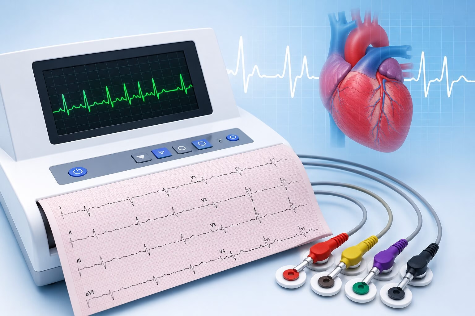
Your heart beats thousands of times every day, and each beat is controlled by electrical signals that help maintain a steady rhythm. When something affects these signals, you may notice symptoms such as a racing heartbeat, palpitations, dizziness, chest discomfort, or shortness of breath. An ECG test is one of the simplest ways for healthcare professionals to check how your heart is beating. An ECG, or electrocardiogram, is a quick, painless, and non-invasive test that records the electrical activity of the heart. It can help identify abnormal heart rhythms and other changes that may require further cardiac evaluation. At Health 24/7, ECG testing is available as part of our cardiac diagnostic services, helping patients receive timely information about their heart health. This guide covers everything you need to know about an ECG test, including why it is performed, how it works, what it can detect, how to prepare, and how to understand your results. Book Your ECG Test Today What Is an ECG (Electrocardiogram) Test? An ECG (electrocardiogram) is a diagnostic test that records the electrical signals produced by your heart as it beats. You may also see it referred to as an EKG, particularly in some countries, but both terms describe the same test. Every heartbeat begins with an electrical impulse. These impulses travel through the heart and cause its chambers to contract in a coordinated pattern, allowing blood to circulate throughout the body. An ECG records these electrical signals and displays them as a series of waves on a monitor or printed report. Healthcare professionals can examine these patterns to evaluate your heart rate, rhythm, and electrical conduction. An ECG may provide information about: Heart rate Heart rhythm Electrical conduction through the heart Possible rhythm abnormalities Changes that may suggest heart strain Certain signs associated with a current or previous heart attack The test does not send electricity into your body. The electrodes simply detect the electrical activity naturally produced by your heart. How Does an ECG Test Work? An ECG uses small adhesive patches called electrodes, which are attached to specific areas of your chest, arms, and legs. Thin wires connect these electrodes to the ECG machine. As your heart beats, the electrodes detect tiny electrical signals and transmit them to the machine. The ECG machine then converts these signals into wave patterns. Different parts of the waveform represent different stages of each heartbeat. Healthcare professionals examine factors such as: How quickly the heart is beating Whether the rhythm is regular How electrical signals travel through the heart Whether certain waveform changes are present A standard resting ECG usually takes only a few minutes to record and causes no pain. Why Is an ECG Test Done? Doctors may recommend an ECG for many reasons, from investigating symptoms to monitoring an existing heart condition. Some of the most common reasons include: Chest Pain or Discomfort Chest pain has many possible causes, ranging from muscle strain and digestive problems to heart-related conditions. An ECG is often one of the first tests performed when a healthcare professional suspects a cardiac cause. However, an ECG alone cannot identify every cause of chest pain, so additional blood tests or imaging may be necessary. Heart Palpitations Palpitations can make your heart feel as though it is racing, pounding, fluttering, or skipping beats. An ECG can help determine whether these sensations are associated with an abnormal heart rhythm. Irregular Heartbeat If your doctor detects an irregular pulse during an examination, an ECG can provide more information about the heart’s rhythm and electrical activity. Shortness of Breath Breathing difficulties can have many causes. When a heart-related problem is suspected, an ECG may be included as part of the initial evaluation. Dizziness or Fainting Sudden dizziness, light-headedness, or fainting can sometimes occur when the heart is beating too quickly, too slowly, or irregularly. An ECG may help identify rhythm abnormalities that could contribute to these symptoms. Monitoring Existing Heart Conditions People diagnosed with certain heart conditions may undergo ECG testing periodically to monitor their heart rhythm and detect changes over time. Before Certain Medical Procedures An ECG may be requested before surgery or another medical procedure, particularly for patients with cardiovascular risk factors or known heart conditions. What Can an ECG Detect? An ECG provides important information about the heart’s electrical activity. However, it should always be interpreted alongside symptoms, medical history, examination findings, and other tests when required. Abnormal Heart Rhythms One of the main uses of an ECG is detecting arrhythmias, which occur when the heart beats too quickly, too slowly, or irregularly. Examples include: Atrial fibrillation Tachycardia Bradycardia Certain conduction abnormalities Some rhythm problems occur intermittently and may not appear during a short resting ECG. In such situations, longer-term heart monitoring may be recommended. Signs of a Heart Attack Certain ECG changes can occur during a heart attack and may help doctors determine which part of the heart is affected. An ECG may also show changes associated with a previous heart attack. However, a normal ECG does not completely rule out a heart attack, so doctors may use additional tests such as cardiac blood tests when clinically necessary. Reduced Blood Flow to the Heart Changes in the ECG waveform can sometimes suggest that part of the heart muscle is not receiving enough blood and oxygen. If symptoms mainly occur during physical activity, additional testing may be required to assess how the heart responds to exercise. Heart Enlargement or Strain Certain ECG patterns may suggest enlargement or increased strain affecting parts of the heart. Further tests, particularly an echocardiogram, may then be recommended to examine the heart’s structure and pumping function in greater detail. Effects of Certain Medications Some medications can affect the heart’s rhythm or electrical conduction. Doctors may use ECG testing to monitor patients taking medicines known to influence cardiac electrical activity. Types of ECG Tests Not every heart rhythm problem appears during a short examination. Different types of ECG monitoring are therefore available depending on the patient’s symptoms. Resting ECG

Open MRI Scan: Benefits, Cost, Procedure, Preparation & Who Should Choose It (Complete Guide)

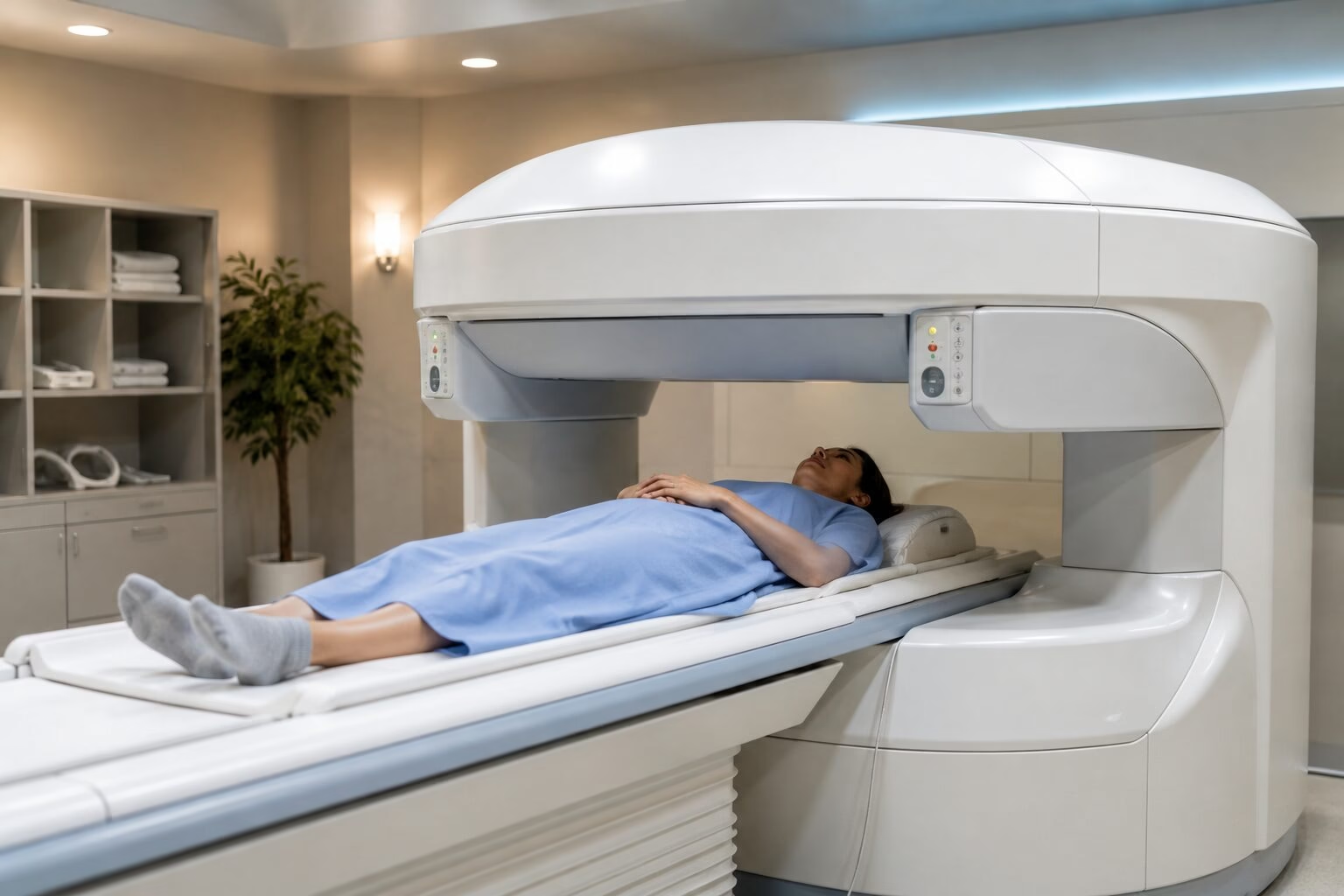
Magnetic Resonance Imaging (MRI) is one of the most advanced diagnostic imaging techniques available today, helping doctors diagnose a wide range of conditions affecting the brain, spine, joints, muscles, and internal organs. While MRI scans provide highly detailed images without using radiation, many patients feel anxious about undergoing the examination because of the enclosed design of traditional MRI machines. For individuals with claustrophobia, anxiety, mobility challenges, or larger body sizes, an Open MRI offers a more comfortable imaging experience while still providing the detailed diagnostic information doctors need for many medical conditions. At Health 24/7, we understand that patient comfort is just as important as diagnostic accuracy. Our MRI services are designed to provide high-quality imaging in a supportive environment, helping patients feel more relaxed throughout the examination. This guide explains everything you need to know about Open MRI scans, including how they work, who may benefit from them, and what to expect during the procedure. Book Your Open MRI Scan Today What Is an Open MRI Scan? An Open MRI scan is a type of Magnetic Resonance Imaging (MRI) that uses powerful magnets and radio waves to create detailed images of the body’s internal structures. Unlike a traditional closed MRI scanner, an Open MRI features a more spacious design with open sides or a wider scanning area, making the experience more comfortable for many patients. Like conventional MRI, Open MRI does not use ionising radiation. Instead, it produces highly detailed images of soft tissues, bones, joints, muscles, ligaments, nerves, and organs using magnetic fields and radiofrequency waves. The open design helps reduce the feeling of being confined, making it a preferred option for patients who may find a standard MRI challenging. Doctors may recommend an Open MRI to evaluate conditions affecting the: Brain Spine Neck Shoulders Knees Hips Ankles Joints Muscles Ligaments Certain abdominal and pelvic structures How Does an Open MRI Scan Work? Although the scanner looks different from a conventional MRI machine, the technology works in a similar way. During the examination: You lie comfortably on a movable scanning table. The table is positioned within the open MRI system. Powerful magnets create a magnetic field around the area being examined. Radio waves temporarily interact with hydrogen atoms in your body. A computer processes the signals to produce detailed cross-sectional images. Unlike X-rays or CT scans, MRI does not expose you to radiation. Instead, it creates exceptionally detailed images of soft tissues that may not be visible with other imaging techniques. Depending on the body part being examined, the scan usually takes between 20 and 60 minutes. Open MRI vs. Traditional MRI Both Open MRI and traditional MRI use the same basic technology to produce detailed images of the body. However, there are important differences in patient comfort and scanner design. Feature Open MRI Traditional MRI Scanner Design Open-sided or wider design Fully enclosed tunnel Patient Comfort Higher Moderate Suitable for Claustrophobic Patients Yes, often preferred May be challenging Image Quality Excellent for many examinations Excellent Scan Time May be slightly longer Usually shorter Best For Patients needing a more comfortable experience Most routine MRI examinations While Open MRI provides excellent diagnostic images for many conditions, certain specialised examinations may still require a high-field traditional MRI system. Your doctor or radiologist will recommend the most appropriate scanner based on your medical condition and the type of images needed. Who Should Consider an Open MRI? Open MRI has been developed to make MRI examinations more comfortable for patients who may struggle with traditional scanners. Patients with Claustrophobia One of the most common reasons patients choose an Open MRI is claustrophobia. The open design reduces the feeling of confinement, helping many patients complete the examination without significant anxiety. Patients with Anxiety Some individuals feel nervous or anxious about remaining inside a traditional MRI scanner for an extended period. The spacious design of an Open MRI often creates a calmer experience, allowing patients to feel more relaxed during the scan. Patients with Larger Body Sizes Open MRI systems typically provide more space around the patient, making them a more comfortable option for individuals with larger body sizes or those who find enclosed scanners restrictive. Elderly Patients Older adults who have difficulty remaining in confined spaces or who require additional comfort during imaging may benefit from an Open MRI examination. Children Some children who need MRI imaging may tolerate an Open MRI better because of its less enclosed design. Depending on the child’s age and ability to remain still, sedation may still be necessary in certain cases. Patients with Limited Mobility Patients with chronic pain, arthritis, spinal disorders, or limited mobility often find it easier to position themselves comfortably within an Open MRI system. What Conditions Can an Open MRI Diagnose? An Open MRI can help diagnose many of the same conditions as a traditional MRI, particularly those involving soft tissues, joints, and the nervous system. Common uses include: Brain Disorders Open MRI may be used to evaluate: Headaches Stroke Brain tumours Multiple sclerosis Seizure disorders Brain injuries Spine Conditions MRI provides detailed images of the cervical, thoracic, and lumbar spine. It helps diagnose: Herniated discs Spinal stenosis Degenerative disc disease Nerve compression Spinal injuries Joint Injuries Doctors frequently request Open MRI scans to evaluate: Knee injuries Shoulder pain Hip disorders Ankle injuries Wrist problems The scan clearly shows cartilage, ligaments, tendons, and surrounding soft tissues. Sports Injuries Athletes often undergo MRI examinations to assess: Ligament tears Muscle injuries Tendon damage Cartilage injuries Joint inflammation Because MRI provides excellent soft tissue contrast, it is considered one of the best imaging techniques for sports medicine. Arthritis and Joint Disorders MRI helps evaluate both inflammatory and degenerative joint diseases by providing detailed images of cartilage, ligaments, bone marrow, and surrounding tissues. Soft Tissue Abnormalities Open MRI can also detect abnormalities involving: Muscles Tendons Ligaments Soft tissue masses Certain pelvic and abdominal structures when clinically indicated By producing detailed, high-resolution images, Open MRI helps healthcare providers diagnose conditions accurately and develop personalised treatment plans. Open MRI Procedure:

X-Ray Scan: Types, Uses, Cost, Procedure, Preparation & Results (Complete Guide)

An X-ray scan is one of the fastest and most widely used diagnostic imaging tests in modern healthcare. Whether you’ve suffered a sports injury, are experiencing persistent chest pain, or need to evaluate a possible fracture, an X-ray provides doctors with valuable information to diagnose a wide range of medical conditions quickly and accurately. Although X-rays have been used in medicine for more than a century, today’s digital X-ray technology offers clearer images, faster reporting, and lower radiation exposure than traditional film-based X-rays. From detecting broken bones and lung infections to identifying joint disorders and certain abdominal conditions, X-rays remain one of the first imaging tests recommended by healthcare professionals. At Health 24/7, we offer advanced digital X-ray services performed by experienced radiologists using modern imaging equipment. This guide explains everything you need to know about X-ray scans, including their uses, types, procedure, preparation, safety, and what to expect after your examination. Book Your X-Ray Scan Today. What Is an X-Ray Scan? An X-ray scan is a diagnostic imaging test that uses a small amount of electromagnetic radiation to create images of the inside of the body. Different tissues absorb X-rays differently. Dense structures such as bones absorb more radiation and appear white on the image, while muscles, fat, and organs appear in varying shades of grey. Air-filled structures, such as the lungs, appear darker. This contrast allows doctors to examine bones, joints, the chest, and certain soft tissues to identify injuries, infections, and other medical conditions. Today, most healthcare facilities use digital X-ray systems, which produce high-quality images almost instantly. Compared with traditional film X-rays, digital imaging offers several advantages, including: Faster image processing Improved image quality Lower radiation exposure Easy electronic storage and sharing Faster diagnosis and reporting Because the procedure is quick, non-invasive, and widely available, X-rays are often the first imaging test recommended when investigating many medical concerns. While X-rays are ideal for evaluating bones and certain chest conditions, they may not always provide enough detail for soft tissues or internal organs. If your doctor requires more detailed images, you can learn more in our MRI Scan Guide, which explains when MRI is the preferred imaging option. How Does an X-Ray Scan Work? An X-ray machine produces a controlled beam of radiation that passes through the body. As the X-rays travel through different tissues, each structure absorbs the radiation differently based on its density. For example: Bones absorb most of the X-rays and appear white. Soft tissues absorb less radiation and appear in shades of grey. Air-filled spaces, such as the lungs, absorb very little radiation and appear black. A digital detector positioned behind the body captures the X-rays that pass through and instantly converts them into detailed digital images. The radiographer may take images from several angles to provide a more complete view of the area being examined. These images are then reviewed by a radiologist, who prepares a report for your referring doctor. The entire process usually takes only a few minutes, making X-rays one of the quickest diagnostic imaging procedures available. Types of X-Ray Scans Different types of X-rays are used to examine specific areas of the body. Your doctor will recommend the most appropriate scan based on your symptoms and medical history. Chest X-Ray A chest X-ray is one of the most frequently performed imaging tests. It helps evaluate: Lungs Heart Airways Ribs Chest cavity Doctors commonly recommend a chest X-ray to investigate: Persistent cough Chest pain Shortness of breath Pneumonia Tuberculosis Lung infections Enlarged heart Fluid around the lungs It is also commonly used during routine health screenings and before certain surgical procedures. Bone X-Ray Bone X-rays provide detailed images of the skeleton and are commonly performed following injuries or accidents. They help diagnose: Bone fractures Stress fractures Bone infections Arthritis Bone tumours Bone deformities Orthopaedic specialists frequently use bone X-rays to monitor healing after fractures or surgery. Spine X-Ray Spine X-rays evaluate the vertebrae and alignment of the spinal column. Depending on your symptoms, your doctor may request: Cervical spine X-ray (neck) Thoracic spine X-ray (upper back) Lumbar spine X-ray (lower back) These scans help identify fractures, spinal curvature, arthritis, degenerative changes, and other spinal abnormalities. Joint X-Ray Joint X-rays examine the bones that form major joints throughout the body. Common joint X-rays include: Knee Shoulder Hip Elbow Wrist Hand Ankle Foot These scans are useful for evaluating joint injuries, arthritis, dislocations, fractures, and age-related wear and tear. Dental X-Ray Dental X-rays allow dentists to examine teeth, jawbones, and surrounding oral structures. Different types include: Bitewing X-rays Periapical X-rays Panoramic dental X-rays (OPG) Routine dental X-rays help detect cavities, impacted teeth, gum disease, bone loss, infections, and other oral health conditions. For more detailed three-dimensional dental imaging, your dentist may recommend a CBCT scan, which provides comprehensive 3D images for implant planning, orthodontics, and complex dental procedures. Abdominal X-Ray Although ultrasound and CT scans are often preferred for abdominal conditions, an abdominal X-ray remains useful in certain situations. It may help identify: Bowel obstruction Kidney stones Swallowed foreign objects Intestinal gas patterns Constipation Your doctor will determine whether an X-ray or another imaging test is more appropriate for your symptoms. Skull X-Ray A skull X-ray examines the bones of the skull and facial structures. Although it has become less common due to advances in CT and MRI imaging, it may still be used in selected cases to evaluate fractures or certain bone abnormalities. When Do You Need an X-Ray? Your healthcare provider may recommend an X-ray whenever a quick assessment of bones, joints, or certain organs is needed. Common reasons include: Bone injuries Suspected fractures Sports injuries Joint pain Persistent back or neck pain Chest pain Chronic cough Shortness of breath Swelling Arthritis Dental pain Suspected lung infections Monitoring healing after surgery or fractures Because X-rays are fast, affordable, and widely available, they often serve as the first step in diagnosing many medical conditions. If your symptoms involve abdominal pain, pregnancy, or blood vessel evaluation, your healthcare provider may recommend an

Ultrasound Scan in Dubai: Types, Cost, Procedure, Preparation & Results (Complete Guide)

An ultrasound scan is one of the most widely used diagnostic imaging tests in modern medicine. It is quick, non-invasive, and safe, making it an essential tool for diagnosing a wide range of medical conditions. From monitoring a healthy pregnancy to evaluating abdominal pain, thyroid disorders, kidney problems, and heart health, ultrasound imaging helps doctors examine internal organs in real time without exposing patients to radiation. In Dubai, ultrasound scans are available at many hospitals and diagnostic centres. However, choosing the right type of ultrasound and understanding how the procedure works can be confusing, especially if it’s your first scan. Patients often have questions about the different types of ultrasound scans, preparation requirements, costs, and how long it takes to receive results. This comprehensive guide explains everything you need to know about ultrasound scans in Dubai, including the different types, how they are performed, when they are recommended, and what you can expect before and after your appointment.If you’re looking for accurate and reliable diagnostic imaging, Health 24/7 offers advanced ultrasound services performed by experienced radiologists using modern imaging technology to support timely diagnosis and treatment. Book Your At-Home Echo Test Today. What Is an Ultrasound Scan? An ultrasound scan, also known as sonography or USG (Ultrasonography), is a medical imaging technique that uses high-frequency sound waves to produce real-time images of organs, tissues, muscles, blood vessels, and other structures inside the body. Unlike CT scans or X-rays, ultrasound imaging does not use ionising radiation, making it one of the safest diagnostic tools available. Because of its excellent safety profile, it is commonly used during pregnancy, in children, and for repeated follow-up examinations. Doctors use ultrasound scans to diagnose medical conditions, monitor ongoing treatments, guide certain medical procedures, and evaluate the health of various organs throughout the body. Some of the most common uses include: Examining abdominal organs Monitoring pregnancy Evaluating pelvic organs Assessing thyroid abnormalities Detecting gallstones and kidney stones Evaluating blood flow through arteries and veins Examining muscles, tendons, and soft tissues Assessing heart function through echocardiography How Does an Ultrasound Scan Work? Ultrasound technology works by sending high-frequency sound waves into the body using a handheld device called a transducer. Before the scan begins, the sonographer applies a clear, water-based gel to the skin. The gel removes tiny air pockets between the skin and the transducer, allowing the sound waves to travel more effectively. As the sound waves pass through the body, they bounce off different tissues and organs. The reflected sound waves are then converted into real-time images displayed on a monitor, allowing the healthcare provider to observe the body’s internal structures immediately. The procedure is painless for most patients, although slight pressure may be felt when the transducer is moved over the area being examined. Because ultrasound produces live images, doctors can also observe organ movement, blood flow, and fetal development in real time. Types of Ultrasound Scans Ultrasound technology is used in many areas of medicine. The type of scan recommended depends on your symptoms and the part of the body being examined. Abdominal Ultrasound An abdominal ultrasound evaluates the organs within the upper abdomen. It can assess the: Liver Gallbladder Pancreas Kidneys Spleen Major blood vessels Doctors commonly recommend an abdominal ultrasound for patients experiencing: Abdominal pain Gallstones Kidney stones Liver disease Unexplained swelling Digestive disorders Pelvic Ultrasound A pelvic ultrasound examines organs within the pelvis. For women, it evaluates: Uterus Ovaries Cervix Fallopian tubes (when visible) Bladder For men, it may assess: Prostate gland Bladder Surrounding pelvic structures Pelvic ultrasounds are commonly performed to investigate pelvic pain, abnormal bleeding, fertility concerns, ovarian cysts, fibroids, or prostate-related conditions. Pregnancy Ultrasound Pregnancy ultrasounds are performed throughout different stages of pregnancy to monitor the health and development of both the mother and baby. Common pregnancy scans include: Dating scan Nuchal translucency (NT) scan Anomaly scan Growth scan Biophysical profile 3D ultrasound 4D ultrasound These scans help monitor fetal growth, detect congenital abnormalities, assess placental position, and evaluate amniotic fluid levels. If you prefer the convenience of receiving pregnancy imaging at home, you can also read our guide on at-home ultrasound for pregnancy, which explains how home ultrasound services work and when they may be appropriate: Breast Ultrasound A breast ultrasound uses sound waves to evaluate breast tissue. It is commonly recommended: After an abnormal mammogram To investigate breast lumps To distinguish between cysts and solid masses During pregnancy or breastfeeding when mammography may not be suitable Breast ultrasound is often used alongside mammography for a more complete breast evaluation. Thyroid Ultrasound A thyroid ultrasound produces detailed images of the thyroid gland located in the neck. Doctors use it to evaluate: Thyroid nodules Enlarged thyroid gland Thyroid cysts Goitre Suspected thyroid cancer Because it is quick and radiation-free, it is one of the preferred imaging tests for thyroid disorders. Neck Ultrasound A neck ultrasound helps examine: Enlarged lymph nodes Salivary glands Neck masses Soft tissue abnormalities It is often recommended when swelling or lumps are detected during a physical examination. Testicular Ultrasound A testicular ultrasound provides detailed images of the testes and surrounding tissues. It may be recommended for: Testicular pain Swelling Trauma Varicocele Hydrocele Suspected testicular tumours The procedure is safe, painless, and highly accurate. Doppler Ultrasound Unlike a standard ultrasound, a Doppler ultrasound evaluates blood flow through arteries and veins. It helps diagnose: Blood clots Varicose veins Peripheral artery disease Carotid artery disease Poor circulation It is also frequently used during pregnancy to assess blood flow between the placenta and the baby. Musculoskeletal Ultrasound Musculoskeletal ultrasound examines: Muscles Tendons Ligaments Joints Soft tissues It is commonly used by sports medicine specialists and orthopaedic doctors to diagnose tendon injuries, muscle tears, inflammation, and joint disorders. Soft Tissue Ultrasound A soft tissue ultrasound evaluates lumps or swellings beneath the skin. It can help identify: Lipomas Cysts Abscesses Enlarged lymph nodes Soft tissue tumours This scan helps determine whether a lump requires further investigation or treatment. Cardiac Ultrasound (Echocardiogram) An echocardiogram is a specialised ultrasound that examines the heart.

Sperm DNA Fragmentation Test: Cost, Normal Range, Results & Importance for Fertility

Sperm DNA Fragmentation Test: Cost, Normal Range, Results & Importance for Fertility

When couples struggle to conceive, the focus often turns to female fertility. However, male factors contribute to nearly half of all infertility cases, making a thorough evaluation of sperm health just as important. While a standard semen analysis provides valuable information about sperm count, movement, and shape, it does not reveal whether the sperm’s genetic material is healthy enough to support successful fertilization and embryo development. This is where the Sperm DNA Fragmentation Test plays a vital role. The test measures the level of DNA damage within sperm cells, offering deeper insights into male fertility that cannot be detected through routine semen analysis alone. It is particularly useful for couples facing unexplained infertility, repeated miscarriages, or unsuccessful IVF cycles. If you are planning to start a family or have experienced fertility challenges, the specialists at Health 24/7 can help you determine whether a Sperm DNA Fragmentation Test is right for you and guide you toward the most appropriate fertility evaluation. Book Your At-Home Echo Test Today. What Is a Sperm DNA Fragmentation Test? A Sperm DNA Fragmentation Test is an advanced laboratory test that evaluates the integrity of the genetic material carried by sperm. Every sperm cell contains DNA that is responsible for passing genetic information to the embryo. When this DNA becomes damaged or fragmented, the sperm may still appear normal under a microscope but may have a reduced ability to achieve a healthy pregnancy. Unlike a routine semen analysis, which focuses on the physical characteristics of sperm, DNA fragmentation testing looks deeper into the quality of the genetic material itself. Understanding DNA Damage in Sperm DNA fragmentation refers to breaks or damage within the DNA strands inside sperm cells. Small amounts of DNA damage can occur naturally and may be repaired by the egg after fertilization. However, when the level of fragmentation is high, it can interfere with fertilization, embryo development, implantation, and the chances of carrying a pregnancy to term. Several factors can contribute to sperm DNA damage, including: Age Smoking and tobacco use Excessive alcohol consumption Obesity Chronic stress Exposure to environmental toxins High fever or infections Varicocele (enlarged veins in the scrotum) Poor lifestyle habits Because many of these factors are preventable or manageable, identifying DNA fragmentation early can help improve fertility outcomes. Why Does It Affect Fertility? Healthy sperm DNA is essential for successful conception and normal embryo development. When DNA fragmentation is elevated, couples may experience: Difficulty conceiving naturally Poor fertilization during IVF or ICSI Slow embryo development Recurrent implantation failure Repeated miscarriages Lower pregnancy success rates Even if a semen analysis appears normal, high DNA fragmentation can still reduce fertility potential. This is why many fertility specialists recommend this advanced test when routine investigations fail to explain infertility. Who Should Consider This Test? A Sperm DNA Fragmentation Test may be recommended for men who: Have unexplained infertility Have experienced repeated IVF or ICSI failure Have had recurrent pregnancy loss with their partner Show normal semen analysis results despite infertility Are over the age of 35 or 40 Have known varicocele Smoke regularly Have diabetes or chronic medical conditions Have been exposed to heat, chemicals, or radiation in the workplace It can also be valuable for couples who want a more comprehensive fertility assessment before beginning assisted reproductive treatment. Why Is a Sperm DNA Fragmentation Test Important? Male fertility is about much more than sperm count. Even when sperm concentration and motility appear healthy, damaged DNA can significantly affect the ability to achieve and maintain a successful pregnancy. Male Infertility Male infertility affects millions of couples worldwide. While conventional semen analysis identifies many common sperm abnormalities, it cannot assess genetic quality. DNA fragmentation testing fills this important gap by identifying hidden sperm damage that may otherwise go unnoticed. Repeated IVF Failure Some couples undergo multiple IVF or ICSI cycles without success despite producing good-quality embryos. One possible explanation is high sperm DNA fragmentation. Research has shown that elevated DNA damage may reduce implantation rates and increase the likelihood of failed fertility treatments. Identifying this issue allows fertility specialists to adjust treatment strategies or recommend interventions that may improve outcomes. Recurrent Miscarriages Pregnancy loss can be emotionally devastating, especially when it happens repeatedly without an obvious cause. Emerging evidence suggests that elevated sperm DNA fragmentation may increase the risk of recurrent miscarriage by affecting embryo development during the earliest stages of pregnancy. Although female factors are often investigated first, assessing male genetic quality can provide valuable additional information. Infertility Despite Normal Semen Analysis A normal semen analysis does not always mean fertility is normal. Traditional semen analysis evaluates: Sperm count Sperm motility Sperm morphology Semen volume However, it cannot determine whether the DNA inside the sperm is intact. For couples with unexplained infertility, DNA fragmentation testing often provides answers when routine fertility investigations appear normal. How Is a Sperm DNA Fragmentation Test Done? The procedure is straightforward and similar to providing a semen sample for routine fertility testing. Sample Collection A semen sample is collected through masturbation into a sterile laboratory container. Patients are usually advised to maintain 2 to 5 days of sexual abstinence before sample collection to ensure accurate results. Many fertility centres request that samples be produced onsite, although some laboratories may allow home collection if strict transport guidelines are followed. If you’re preparing for semen testing, our guide on common semen analysis preparation mistakes can help you avoid errors that may affect your results: Laboratory Testing Methods After collection, the sample is processed using specialised laboratory techniques. Common testing methods include: Halo Test (Sperm Chromatin Dispersion Test) This method measures the integrity of sperm DNA by examining the halo that forms around sperm cells after laboratory processing. It is widely used because it is reliable and relatively simple. TUNEL Assay The TUNEL method directly identifies fragmented DNA by labeling damaged DNA strands. It is considered one of the most accurate laboratory techniques for assessing DNA fragmentation. SCSA (Sperm Chromatin Structure Assay) SCSA uses advanced flow cytometry to evaluate sperm

GI-MAP vs. Standard Stool Test: Which Gut Health Test Do You Actually Need?

Gut health has become one of the biggest topics in modern healthcare, and for good reason. Your digestive system does far more than break down food. It influences your immune system, energy levels, mental well-being, metabolism, and overall quality of life. As awareness grows, more people are seeking answers for persistent digestive symptoms such as bloating, abdominal pain, irregular bowel movements, and food sensitivities. The challenge is that many people try to manage these symptoms through trial and error. They change diets, take supplements, or eliminate foods without understanding what’s actually happening inside their gut. This often leads to wasted time, unnecessary expenses, and ongoing frustration. One of the biggest sources of confusion is choosing the right diagnostic test. Should you undergo a standard stool test ordered by your doctor, or would a more advanced GI-MAP test provide the answers you’re looking for? The answer depends on your symptoms and health goals. A standard stool test is best for detecting acute infections and urgent digestive illnesses, while a GI-MAP test is designed for people experiencing chronic digestive issues who need a deeper understanding of their gut microbiome. Let’s explore how these tests differ and which one may be the right choice for you. Connect with our team on WhatsApp. What Is a Standard Stool Test? A standard stool test is one of the most commonly requested laboratory investigations for digestive complaints. It is typically recommended when a healthcare provider suspects an infection, gastrointestinal bleeding, or a parasite. Depending on the clinical situation, the laboratory may perform stool culture, microscopy, or other routine analyses to identify common pathogens or abnormalities. What It Measures & Detects A standard stool test can help identify: Bacterial infections such as E. coli, Salmonella, and Shigella Certain parasites and intestinal worms Blood in the stool Signs of inflammation Some viral infections (depending on the test ordered) Because it focuses primarily on disease-causing organisms, it is highly effective for diagnosing acute gastrointestinal infections. Best Used For A standard stool test is usually recommended if you experience: Sudden or severe diarrhea Food poisoning Fever accompanied by digestive symptoms Bloody stools Recent travel-related infections Routine colorectal screening when occult blood testing is required For these situations, standard stool testing remains the first-line diagnostic tool because it quickly identifies infections that require immediate treatment. What Is a GI-MAP Test? (The Functional Deep-Dive) Unlike traditional stool analysis, the GI-MAP (Gastrointestinal Microbial Assay Plus) is an advanced DNA-based stool test that evaluates the overall health of your gut microbiome. Using quantitative Polymerase Chain Reaction (qPCR) technology, the GI-MAP identifies microbial DNA present in your stool sample. Rather than looking only for harmful infections, it provides a detailed picture of the beneficial bacteria, harmful microorganisms, digestive markers, immune function, and inflammation within your digestive tract. If you’re unfamiliar with how this advanced assessment works, read our GI-MAP Test Dubai Guide What It Measures & Detects A GI-MAP test provides comprehensive information about: Beneficial gut bacteria Opportunistic bacteria Harmful pathogens Helicobacter pylori (H. pylori) Yeast and Candida overgrowth Parasites Viruses Digestive enzyme activity Inflammatory markers Leaky gut (intestinal permeability) indicators Immune response markers Instead of simply identifying whether an infection exists, the test helps explain why digestive symptoms may be occurring. Best Used For A GI-MAP test may be recommended for people experiencing: Chronic bloating Irritable Bowel Syndrome (IBS) Constipation or diarrhea lasting several weeks Food sensitivities Suspected leaky gut Autoimmune conditions Persistent fatigue Skin conditions linked to gut health Recurrent digestive discomfort despite normal routine tests If you’ve been struggling with ongoing digestive symptoms and routine investigations haven’t provided answers, learn who should consider a GI-MAP test. Key Differences: Technology, Cost, and Insights Although both tests involve analyzing a stool sample, they are designed for different clinical purposes. Culture vs. DNA Technology A standard stool test usually relies on stool culture or microscopic examination. While effective for identifying many common pathogens, it can sometimes miss organisms that are difficult to culture or present in low numbers. The GI-MAP uses advanced qPCR technology to detect microbial DNA directly. This enables it to identify a wider range of microorganisms with greater sensitivity, including organisms that traditional culture methods may overlook. Cost and Insurance Coverage Standard stool tests are commonly covered by health insurance when medically indicated, making them an affordable option for diagnosing acute gastrointestinal illnesses. A GI-MAP test is a specialized functional medicine assessment. Coverage varies depending on your insurance provider, and many patients choose to pay privately for the additional insights it provides. While the initial investment may be higher, it can help identify underlying causes of chronic digestive symptoms that would otherwise remain undiagnosed.To learn more about the technology, cost, and insights of gut microbiome testing, read Gut Microbiome Test in Dubai & UAE: Price, Process & What It Reveals. Actionable Insights for Long-Term Wellness One of the biggest differences lies in the depth of information. A standard stool test answers a straightforward question: “Do you currently have an infection or other obvious abnormality?” A GI-MAP answers a much broader question: “What is happening throughout your gut ecosystem, and what could be contributing to your symptoms?” The results can help healthcare providers create personalized plans involving nutrition, probiotics, antimicrobial therapy, digestive support, and lifestyle modifications. The Verdict: Which Test Should You Get? The right test depends on why you’re being evaluated. Feature Standard Stool Test GI-MAP Test Purpose Detect acute infections Comprehensive gut microbiome analysis Technology Culture & microscopy DNA-based qPCR Detects bacterial infections Detects parasites (more comprehensive) Evaluates beneficial bacteria ✘ Assesses gut microbiome balance ✘ Detects Candida & yeast Limited Measures inflammation markers Limited Evaluates digestive function ✘ Best for chronic digestive symptoms Limited Choose a Standard Stool Test If: A traditional stool test is generally the right choice if you: Have sudden diarrhea Suspect food poisoning Have a fever with digestive symptoms Need infection screening Require routine medical investigations Choose a GI-MAP Test If: A GI-MAP test may be the better option if: Your digestive symptoms have lasted for months Previous

Echo vs ECG

Echo vs ECG: Differences, Uses, and Which Heart Test You Need

Heart tests play an important role in detecting and monitoring various cardiac conditions. Among the most commonly recommended heart tests are the ECG and Echo, but they serve different purposes. An ECG records the heart’s electrical activity, while an Echo creates real time images of the heart’s structure and function. Take a look at how these tests differ, what they can detect, and which one may be recommended based on your symptoms and heart health needs. What Is an ECG Test? An ECG, also called an Electrocardiogram, is a quick and non invasive heart test that records the electrical signals controlling each heartbeat. Small electrodes are placed on the chest, arms, and legs to capture electrical activity and display it as a graph for analysis. This test helps detect arrhythmias, atrial fibrillation, abnormal heart rate, conduction abnormalities, and signs of a current or previous heart attack. An ECG can also reveal heart changes linked to electrolyte imbalances and other conditions affecting the heart’s electrical system, making it one of the most commonly used cardiac screening tests. What Is an Echo Test? An Echo, or Echocardiogram, is an ultrasound based heart test that uses sound waves to create moving images of the heart. It allows doctors to examine heart chambers, valves, blood flow, and pumping function without surgery or radiation exposure. An Echo helps diagnose heart valve disease, heart failure, enlarged heart chambers, congenital heart defects, cardiomyopathy, blood flow abnormalities, and pericardial effusion. By showing detailed images of heart structures, it provides valuable information about how effectively the heart is working and whether any physical abnormalities are present. What Is the Main Difference Between Echo and ECG? The main difference between Echo and ECG is that an ECG records the heart’s electrical activity, while an Echo creates ultrasound images of the heart’s structure and function. ECG is mainly used to identify rhythm related issues, whereas Echo helps evaluate heart valves, chambers, blood flow, and pumping performance. Feature ECG Echo Purpose Assess electrical activity Assess structure and function Technology Used Electrodes and electrical recording Ultrasound imaging What It Measures Heart rhythm and electrical signals Heart chambers, valves, blood flow Procedure Duration 5 to 10 minutes 20 to 45 minutes Radiation Exposure None None Detects Rhythm Problems Yes Limited Detects Valve Disorders No Yes Detects Structural Abnormalities Limited Yes Detects Pumping Function No Yes Typical Use Cases Arrhythmias, chest pain, palpitations Valve disease, heart failure, murmurs Book Your At-Home Echo Test Today. When Is an ECG Recommended? An ECG is commonly recommended when doctors need to evaluate the heart’s electrical activity, investigate symptoms, or assess cardiovascular risk. It is often one of the first heart tests performed during cardiac evaluation. Chest Pain: Helps identify signs of reduced blood supply to the heart, previous heart attacks, or electrical changes that may explain chest discomfort. Palpitations: Useful for detecting irregular heart rhythms, rapid heartbeats, and rhythm disturbances that may cause a fluttering or racing sensation. Dizziness or Fainting: Can reveal abnormal heart rhythms or conduction problems that may reduce blood flow to the brain and cause fainting episodes. High Blood Pressure Monitoring: May detect heart strain or electrical changes associated with long term uncontrolled hypertension. Pre Surgical Heart Assessment: Frequently performed before surgery to identify hidden heart rhythm abnormalities and assess overall cardiac status. When Is an Echo Recommended? An Echo is recommended when doctors need detailed information about the heart’s structure, valves, blood flow, and pumping ability. It is often used when symptoms suggest a physical heart abnormality. Heart Murmurs: Helps identify valve narrowing, valve leakage, or structural abnormalities causing unusual heart sounds. Shortness of Breath: Can reveal heart failure, weakened heart muscles, valve disease, or other conditions affecting blood circulation. Suspected Heart Failure: Evaluates heart pumping strength, chamber size, and overall cardiac function to support diagnosis and treatment planning. Valve Disease Monitoring: Provides detailed images that help track progression of valve narrowing or leakage over time. Follow Up After Heart Attack: Assesses heart muscle damage, pumping performance, and possible complications after a cardiac event. Can an Echo Detect Problems That an ECG Cannot? Yes. An Echo can detect several heart conditions that may not appear clearly on an ECG. Because it creates real time images of the heart, it can identify valve abnormalities, enlarged heart chambers, weakened heart muscles, congenital heart defects, and fluid around the heart. For example, a patient with severe valve leakage may have a relatively normal ECG, while an Echo can directly show the affected valve and blood flow changes. This makes Echocardiography particularly valuable for evaluating structural heart disease. Can an ECG Detect Problems That an Echo Cannot? Yes. An ECG is particularly effective at detecting rhythm disorders and electrical conduction abnormalities that may not be visible on an Echo. It can identify atrial fibrillation, silent arrhythmias, heart blocks, and acute electrical changes associated with heart attacks. Since an ECG records the heart’s electrical signals in real time, it is often the preferred first test when symptoms such as palpitations, dizziness, or sudden irregular heartbeats are present. Do You Need Both Echo and ECG? In some situations, one test may provide enough information. For example, an ECG may be sufficient when evaluating a suspected rhythm disorder, while an Echo may be enough when assessing a heart murmur or suspected valve problem. The choice depends on symptoms, medical history, and clinical findings. Many cardiac conditions require both tests because they provide complementary information. ECG evaluates electrical activity, while Echo assesses structure and function. Cardiologists commonly recommend both tests for chest pain, heart failure symptoms, unexplained shortness of breath, or suspected cardiovascular disease to achieve a more complete and accurate diagnosis. Which Test Is More Accurate for Heart Problems? Which Test Is More Suitable for Different Heart Conditions? Echo and ECG provide different insights into heart health. The most suitable test depends on the condition being investigated, as each evaluates a different aspect of how the heart functions. Suitable Test for Rhythm Disorders An ECG is usually the first test used to

Quadruple Marker Test

Quadruple Marker Test Cost in Dubai: Price, Normal Range, Purpose

Pregnancy screening tests help doctors assess a baby’s health before birth and identify potential concerns early. The Quadruple Marker Test is an important prenatal screening test performed during the second trimester to estimate the risk of certain chromosomal abnormalities and birth defects. Read on to know the quadruple marker test cost in Dubai, what the test checks, who should consider it, and how it supports early pregnancy care. What Is a Quadruple Marker Test The Quadruple Marker Test is a maternal blood test performed during the second trimester of pregnancy. It evaluates four substances in the mother’s blood to estimate the risk of chromosomal abnormalities and certain birth defects. As part of prenatal screening, it helps doctors determine whether additional testing may be beneficial. What Does a Quadruple Marker Test Check The test checks the risk of conditions such as Down syndrome, Edwards syndrome, and neural tube defects. It analyzes Alpha Fetoprotein, Human Chorionic Gonadotropin, Unconjugated Estriol, and Inhibin A levels together with maternal age and pregnancy details to calculate risk estimates. Why Is It Called a Quadruple Marker Test The name comes from the four biological markers measured during testing. Each marker provides different information about fetal development and pregnancy health. Combining all four markers improves screening accuracy and helps create a more reliable risk assessment. Is It a Screening Test or Diagnostic Test The Quadruple Marker Test is a screening test rather than a diagnostic test. It estimates the probability of chromosomal abnormalities but cannot confirm whether a baby has a specific condition. Further testing may be recommended if results indicate increased risk. How Does It Help During Pregnancy The test provides valuable information during the second trimester and helps identify pregnancies that may require additional monitoring. Early risk assessment supports informed medical decisions and allows healthcare providers to recommend appropriate follow up screening or diagnostic procedures.   Book Your Quadruple Marker Test Today. Quadruple Marker Test Cost in Dubai The cost of a Quadruple Marker Test in Dubai varies based on the healthcare provider, testing technology, and additional services included in the package. Most diagnostic centers and hospitals offer this screening as part of pregnancy care services. Average Quadruple Marker Test Price in Dubai The average quadruple marker test cost in Dubai typically ranges from AED 350 to AED 900. Pricing may vary depending on the laboratory, specialist involvement, reporting speed, and package inclusions. What Affects Quadruple Marker Test Cost? Advanced laboratory technology may increase testing costs. Specialist consultations can add to package pricing. Additional scans may be included in packages. Hospital facilities often charge higher testing fees. Diagnostic centers may offer more affordable rates. Faster report delivery can affect overall pricing. Promotional healthcare packages may reduce costs. Does Insurance Cover the Test in Dubai? Insurance coverage depends on the provider and policy terms. Some health insurance plans may cover prenatal screening when medically recommended, while others may require partial payment or offer limited maternity related benefits. Quadruple Marker Test Cost Across Dubai Healthcare Providers Pricing can vary significantly across Dubai based on the type of healthcare facility, technology used, specialist involvement, and package inclusions. Comparing available options helps expectant mothers find the right balance between affordability, convenience, and quality prenatal screening services. Private Hospitals vs Diagnostic Centers Private hospitals generally charge between AED 550 and AED 900 for a Quadruple Marker Test, especially when specialist consultations and maternity services are included. Diagnostic centers often offer the same screening test for AED 350 to AED 700, making them a more affordable choice for routine prenatal screening while maintaining reliable laboratory standards. Standalone Test vs Pregnancy Screening Package A standalone Quadruple Marker Test typically costs between AED 350 and AED 700. Pregnancy screening packages may range from AED 700 to AED 1,500 or more, depending on whether they include specialist consultations, ultrasound scans, blood investigations, fetal assessments, or detailed pregnancy monitoring services. Home Sample Collection Charges Many diagnostic providers offer home sample collection for added convenience. Charges may range from free service promotions to AED 50 to AED 150 depending on the location, appointment timing, and provider policies. Some healthcare platforms include complimentary home sample collection as part of promotional screening packages. How to Choose a Reliable Testing Center When comparing providers, consider the following factors before booking: Accredited laboratory with established quality control standards. Experienced pathology and prenatal screening specialists available. Advanced laboratory equipment for accurate testing results. Transparent pricing without hidden package charges. Fast report turnaround and digital report access. Positive patient reviews and healthcare reputation. Convenient location or home collection availability. Where Can I Get a Quadruple Marker Test in Dubai? The Quadruple Marker Test is available through hospitals, maternity clinics, and diagnostic centers across Dubai. Many healthcare providers also partner with medical booking platforms that allow patients to compare pricing, locations, and available appointment slots. Diagnostic Centers Offering Prenatal Screening Diagnostic centers are among the most popular options for prenatal screening because they often provide competitive pricing, shorter waiting times, online booking facilities, home sample collection services, and multiple branch locations throughout Dubai. Hospital Based Maternal Screening Services Hospital based maternal screening services offer integrated pregnancy care under one roof. Patients may benefit from consultations with obstetricians, maternal fetal medicine specialists, ultrasound services, and laboratory testing during the same visit, although costs are generally higher. How to Book a Quadruple Marker Test Booking a Quadruple Marker Test is usually simple and can be completed through several methods: Contact a hospital or diagnostic center directly. Book through the provider’s official website. Schedule through healthcare mobile applications. Use diagnostic booking platforms for comparisons. Visit the facility and book in person. Request home sample collection if available. Confirm pregnancy week before finalizing appointment. Benefits of Booking Through Healthcare Platforms Healthcare booking platforms can provide several advantages compared to direct booking: Compare prices across multiple diagnostic providers. Access exclusive discounts and promotional offers. Select preferred location based on convenience. View available appointment slots instantly. Book home sample collection services easily. Access patient reviews before booking. Receive digital

triple marker test

What Does a Triple Marker Test Tell You About Your Pregnancy?

Pregnancy involves several screening tests that help doctors assess your baby’s development and identify potential risks early. One such test is the triple marker test, a commonly recommended prenatal screening performed during the second trimester. It provides important information about the likelihood of certain chromosomal conditions and birth defects. Read on to know what the triple marker test reveals, when it is performed, and how the results may influence your pregnancy care. What Is a Triple Marker Test? The triple marker test is a prenatal blood test used during pregnancy to estimate the risk of specific genetic conditions and neural tube defects in a developing baby. It is also known as a triple marker screening test or triple screen test. Unlike diagnostic tests that confirm a condition, this screening test helps identify whether further evaluation may be needed. The test analyzes three substances present in the mother’s blood. The levels of these markers, combined with factors such as maternal age and gestational age, help calculate the likelihood of certain fetal abnormalities. What Does the Triple Marker Test Measure? The triple marker blood test measures three important substances that are naturally produced during pregnancy. These markers provide valuable information about fetal development and pregnancy health. Alpha Fetoprotein (AFP) is produced mainly by the developing baby. Human Chorionic Gonadotropin (hCG) is produced by the placenta. Unconjugated Estriol (uE3) is produced by both the placenta and fetus. AFP levels may indicate possible neural tube defects. hCG levels can help assess chromosomal abnormality risks. Estriol levels reflect fetal and placental wellbeing. Combined marker levels provide a risk estimate. Results are interpreted alongside maternal health information. What Conditions Can the Test Screen For? The triple marker test helps screen for several conditions that may affect fetal development. It does not diagnose these conditions but identifies pregnancies that may require additional evaluation. Down syndrome, also known as Trisomy 21. Edwards syndrome, also known as Trisomy 18. Neural tube defects affecting the brain and spine. Spina bifida, involving incomplete spinal development. Anencephaly, involving incomplete brain formation. Certain chromosomal abnormalities linked to fetal growth issues. Pregnancy complications associated with abnormal marker levels. Increased risk pregnancies requiring further screening. Book Your Triple Marker Test Today. When Is the Triple Marker Test Performed During Pregnancy? Timing plays an important role in obtaining reliable results from the triple marker screening test. The test is typically performed during the second trimester when the marker levels are most useful for risk assessment. Doctors carefully calculate gestational age before ordering the test because marker levels naturally change throughout pregnancy. Testing outside the recommended period can affect result accuracy. Recommended Weeks for Testing The triple marker test is usually scheduled during a specific period of pregnancy to maximize accuracy and improve risk estimation. Commonly performed between 15 and 20 weeks of pregnancy. Best accuracy is often achieved between 16 and 18 weeks. Usually recommended during routine second trimester care. Performed after first trimester screening when applicable. Helps assess fetal development during a critical stage. Supports early identification of potential abnormalities. Provides time for additional testing if needed. Often combined with ultrasound findings for assessment. Why Timing Affects Accuracy The concentration of AFP, hCG, and estriol changes throughout pregnancy. Accurate pregnancy dating is therefore essential when interpreting results. Marker levels rise and fall at different stages. Incorrect gestational age may alter risk calculations. Early testing may produce unreliable estimates. Late testing may reduce predictive value. Ultrasound dating improves result interpretation. Accurate timing reduces false positive findings. Proper scheduling improves screening performance. Doctors use timing to compare results with expected ranges. Who May Be Advised to Have a Triple Marker Test? The triple marker test is offered to many pregnant women as part of routine prenatal screening. In some cases, doctors may place greater emphasis on this test because of specific risk factors identified during pregnancy. The screening helps healthcare providers gather additional information about fetal health and determine whether more advanced testing should be considered. Pregnant women aged 35 years or older. Women with a family history of genetic disorders. Previous pregnancy affected by chromosomal abnormalities. Abnormal findings during first trimester screening. Pregnancies with uncertain genetic risk factors. Women seeking additional prenatal screening information. High risk pregnancies requiring closer monitoring. Patients considering further genetic testing options. Compared with diagnostic procedures such as amniocentesis, the triple marker test is non invasive and involves only a blood sample. It may also be used when patients seek additional screening information before considering advanced prenatal diagnostic procedures. How Is the Triple Marker Test Done? The triple marker test is a simple blood test that can usually be completed within a few minutes. It does not require surgery, hospital admission, or invasive procedures, making it a routine part of prenatal care for many women. Healthcare providers combine laboratory results with pregnancy details to calculate the likelihood of specific fetal conditions. A doctor reviews pregnancy history and risk factors. Gestational age is confirmed using medical records or ultrasound. A blood sample is collected from the mother’s arm. The sample is sent to a laboratory for analysis. AFP, hCG, and estriol levels are measured. Maternal age and pregnancy details are included. Computer based calculations generate risk estimates. Results are reviewed by the healthcare provider. Most women do not need to fast before the test. Normal daily activities can generally continue before and after blood sample collection. The procedure poses no direct risk to the developing baby because only the mother’s blood is analyzed. Triple Marker Test Results and What Happens Next Receiving a triple marker test result can be stressful for some parents, especially when they are unsure how the findings should be interpreted. It is important to remember that this prenatal screening test estimates risk and does not confirm whether a baby has a specific condition. Doctors evaluate the results together with maternal age, gestational age, medical history, and ultrasound findings before recommending any additional steps. How Results Are Interpreted The report generated after a triple marker test provides a calculated

Penile Doppler

What to Expect During a Penile Doppler Examination

Erectile dysfunction can occur for many reasons, including blood flow problems, nerve related conditions, hormonal imbalances, and lifestyle factors. When doctors need a detailed assessment of penile circulation, they may recommend a penile Doppler examination. This specialized test helps evaluate how blood flows through the penile arteries and veins and can provide valuable information for diagnosis and treatment planning.If your doctor has recommended a penile Doppler test, knowing what happens before, during, and after the procedure can help you feel more comfortable and prepared. Why a Penile Doppler Examination May Be Recommended A penile Doppler ultrasound is commonly performed when a patient experiences ongoing erectile difficulties and the exact cause is not clear. The test helps doctors assess whether blood flow issues are contributing to erectile dysfunction. Symptoms That Often Lead to Further Evaluation Men may be advised to undergo a penile Doppler examination if they experience: Difficulty achieving an erection Difficulty maintaining an erection during intercourse Reduced penile rigidity Inconsistent erectile performance Penile curvature or structural concerns Erectile dysfunction that does not improve with standard treatments Conditions That Can Affect Penile Blood Flow Several medical conditions can affect circulation and increase the risk of vascular erectile dysfunction, including: Diabetes High blood pressure High cholesterol Cardiovascular disease Obesity Smoking related vascular damage Previous pelvic injury or surgery   Book Your Doppler Ultrasound Today. What Happens During a Penile Doppler Examination A penile Doppler procedure is generally performed by an experienced urologist, andrologist, or diagnostic imaging specialist. The examination is usually completed within 30 to 60 minutes. Initial Assessment Before Imaging Before the scan begins, the healthcare provider may: Review your medical history Discuss erectile dysfunction symptoms Review medications and previous treatments Perform a brief physical examination Explain the procedure and answer any questions Ultrasound and Blood Flow Measurement Process The examination typically includes the following steps: Ultrasound gel is applied to the penis Baseline ultrasound images are obtained A medication may be administered through a small injection to encourage an erection Blood flow measurements are recorded using Doppler ultrasound technology Additional images are taken during different stages of the erection One of the advantages of a penile Doppler ultrasound is that it evaluates both arterial blood supply and venous function. This allows doctors to identify whether erectile dysfunction is related to reduced blood inflow, excessive blood outflow, or a combination of factors. How to Prepare for the Appointment Most patients require very little preparation before a penile Doppler test. However, a few simple steps can help ensure a smooth experience. Before your appointment: Inform your doctor about all medications you are taking Share details about any existing medical conditions Bring previous medical records if available Follow any instructions provided by the clinic Wear comfortable clothing Penile Doppler Test Cost in Dubai The cost of a penile Doppler examination can vary depending on the healthcare facility, specialist involvement, and diagnostic requirements. With trusted providers like health24/7, the Penile Doppler Examination is available for AED 899, which generally includes specialist assessment, penile Doppler ultrasound imaging, detailed blood flow evaluation, diagnostic reporting, and clinical recommendations based on the findings. This comprehensive evaluation helps identify vascular causes of erectile dysfunction and supports accurate treatment planning. Results, Next Steps, and Treatment Planning After the examination, the collected images and blood flow measurements are carefully reviewed. The results help doctors determine whether erectile dysfunction is linked to vascular causes or other contributing factors. The findings may indicate: Normal penile blood flow Reduced arterial blood supply Venous leakage Structural abnormalities Signs of penile vascular disease Based on the results, the doctor may recommend: Lifestyle modifications Medication therapy Hormonal evaluation Further diagnostic testing Regenerative treatments Surgical options when clinically appropriate A Valuable Step Toward Accurate Diagnosis A penile Doppler examination is a safe and effective diagnostic procedure that helps identify blood flow related causes of erectile dysfunction. By evaluating the function of penile arteries and veins, the test provides detailed information that cannot be obtained through routine examinations alone. For men experiencing ongoing erectile difficulties, a penile Doppler ultrasound can play an important role in identifying the underlying cause and guiding appropriate treatment decisions. Early evaluation and accurate diagnosis often lead to better treatment outcomes and improved quality of life. Safe and Affordable Penile Doppler Examination in Dubai HEALTH24/7 provides advanced Penile Doppler Examination services by offering access to the nearest diagnostic facility across Dubai. Through our network of trusted diagnostic centers and 15+ years experienced radiologists, we ensure safe, accurate, and reliable diagnostics. Patients benefit from affordable pricing, advanced ultrasound technology, experienced healthcare professionals, fast appointment scheduling, and detailed reporting. Our commitment to quality care helps support accurate diagnosis and personalized treatment planning in a comfortable clinical environment. Frequently Asked Questions Is a penile Doppler examination painful? Most of the procedure is painless. Some patients may experience mild discomfort from the small injection used to stimulate blood flow during the test. How long does a penile Doppler test take? A penile Doppler examination generally takes between 30 and 60 minutes, depending on the individual assessment requirements. Do I need an erection during the examination? Yes. A medication may be administered to help achieve an erection so blood flow measurements can be accurately evaluated. Can a penile Doppler test identify the cause of erectile dysfunction? The test is highly effective in identifying vascular causes of erectile dysfunction and helps doctors determine whether blood flow abnormalities are contributing to symptoms. Is penile Doppler ultrasound safe? Yes. The procedure uses ultrasound technology and does not involve radiation exposure, making it a safe diagnostic imaging technique. Who should undergo a penile Doppler examination? Men experiencing erectile dysfunction, suspected vascular abnormalities, Peyronie’s disease, or unexplained erectile difficulties may benefit from this examination. Can I return to normal activities after the test? Most patients can return to their normal daily activities shortly after the procedure unless otherwise advised by their healthcare provider. Book Your At-Home Ultrasound Today.