Heart tests play an important role in detecting and monitoring various cardiac conditions. Among the most commonly recommended heart tests are the ECG and Echo, but they serve different purposes. An ECG records the heart’s electrical activity, while an Echo creates real time images of the heart’s structure and function. Take a look at how these tests differ, what they can detect, and which one may be recommended based on your symptoms and heart health needs.
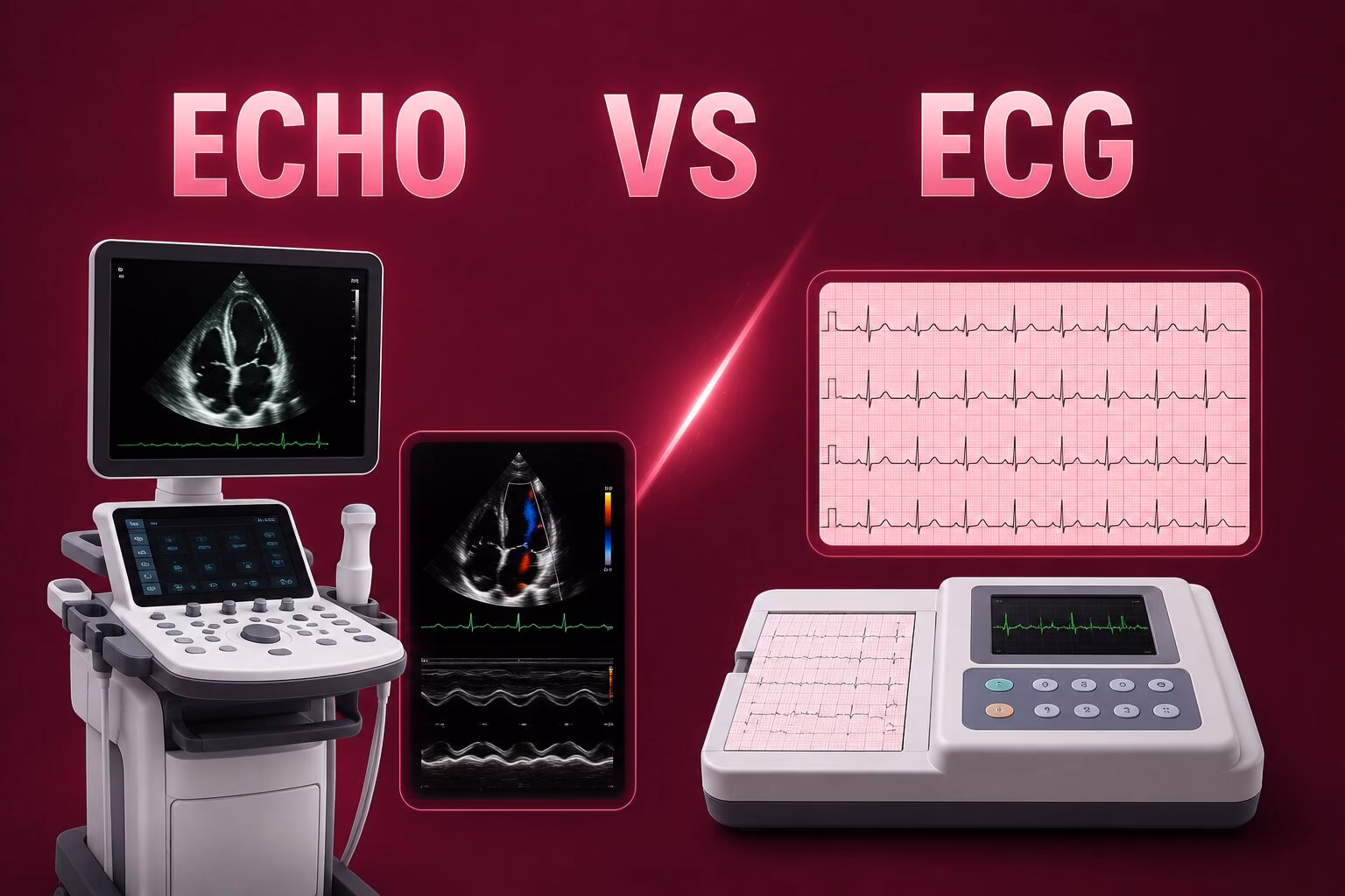
What Is an ECG Test?
An ECG, also called an Electrocardiogram, is a quick and non invasive heart test that records the electrical signals controlling each heartbeat. Small electrodes are placed on the chest, arms, and legs to capture electrical activity and display it as a graph for analysis.
This test helps detect arrhythmias, atrial fibrillation, abnormal heart rate, conduction abnormalities, and signs of a current or previous heart attack. An ECG can also reveal heart changes linked to electrolyte imbalances and other conditions affecting the heart’s electrical system, making it one of the most commonly used cardiac screening tests.
What Is an Echo Test?
An Echo, or Echocardiogram, is an ultrasound based heart test that uses sound waves to create moving images of the heart. It allows doctors to examine heart chambers, valves, blood flow, and pumping function without surgery or radiation exposure.
An Echo helps diagnose heart valve disease, heart failure, enlarged heart chambers, congenital heart defects, cardiomyopathy, blood flow abnormalities, and pericardial effusion. By showing detailed images of heart structures, it provides valuable information about how effectively the heart is working and whether any physical abnormalities are present.
What Is the Main Difference Between Echo and ECG?
The main difference between Echo and ECG is that an ECG records the heart’s electrical activity, while an Echo creates ultrasound images of the heart’s structure and function. ECG is mainly used to identify rhythm related issues, whereas Echo helps evaluate heart valves, chambers, blood flow, and pumping performance.
Feature | ECG | Echo |
Purpose | Assess electrical activity | Assess structure and function |
Technology Used | Electrodes and electrical recording | Ultrasound imaging |
What It Measures | Heart rhythm and electrical signals | Heart chambers, valves, blood flow |
Procedure Duration | 5 to 10 minutes | 20 to 45 minutes |
Radiation Exposure | None | None |
Detects Rhythm Problems | Yes | Limited |
Detects Valve Disorders | No | Yes |
Detects Structural Abnormalities | Limited | Yes |
Detects Pumping Function | No | Yes |
Typical Use Cases | Arrhythmias, chest pain, palpitations | Valve disease, heart failure, murmurs |
When Is an ECG Recommended?
An ECG is commonly recommended when doctors need to evaluate the heart’s electrical activity, investigate symptoms, or assess cardiovascular risk. It is often one of the first heart tests performed during cardiac evaluation.
Chest Pain: Helps identify signs of reduced blood supply to the heart, previous heart attacks, or electrical changes that may explain chest discomfort.
Palpitations: Useful for detecting irregular heart rhythms, rapid heartbeats, and rhythm disturbances that may cause a fluttering or racing sensation.
Dizziness or Fainting: Can reveal abnormal heart rhythms or conduction problems that may reduce blood flow to the brain and cause fainting episodes.
High Blood Pressure Monitoring: May detect heart strain or electrical changes associated with long term uncontrolled hypertension.
Pre Surgical Heart Assessment: Frequently performed before surgery to identify hidden heart rhythm abnormalities and assess overall cardiac status.
When Is an Echo Recommended?
An Echo is recommended when doctors need detailed information about the heart’s structure, valves, blood flow, and pumping ability. It is often used when symptoms suggest a physical heart abnormality.
Heart Murmurs: Helps identify valve narrowing, valve leakage, or structural abnormalities causing unusual heart sounds.
Shortness of Breath: Can reveal heart failure, weakened heart muscles, valve disease, or other conditions affecting blood circulation.
Suspected Heart Failure: Evaluates heart pumping strength, chamber size, and overall cardiac function to support diagnosis and treatment planning.
Valve Disease Monitoring: Provides detailed images that help track progression of valve narrowing or leakage over time.
Follow Up After Heart Attack: Assesses heart muscle damage, pumping performance, and possible complications after a cardiac event.
Can an Echo Detect Problems That an ECG Cannot?
Yes. An Echo can detect several heart conditions that may not appear clearly on an ECG. Because it creates real time images of the heart, it can identify valve abnormalities, enlarged heart chambers, weakened heart muscles, congenital heart defects, and fluid around the heart. For example, a patient with severe valve leakage may have a relatively normal ECG, while an Echo can directly show the affected valve and blood flow changes. This makes Echocardiography particularly valuable for evaluating structural heart disease.
Can an ECG Detect Problems That an Echo Cannot?
Yes. An ECG is particularly effective at detecting rhythm disorders and electrical conduction abnormalities that may not be visible on an Echo. It can identify atrial fibrillation, silent arrhythmias, heart blocks, and acute electrical changes associated with heart attacks. Since an ECG records the heart’s electrical signals in real time, it is often the preferred first test when symptoms such as palpitations, dizziness, or sudden irregular heartbeats are present.
Do You Need Both Echo and ECG?
In some situations, one test may provide enough information. For example, an ECG may be sufficient when evaluating a suspected rhythm disorder, while an Echo may be enough when assessing a heart murmur or suspected valve problem. The choice depends on symptoms, medical history, and clinical findings.
Many cardiac conditions require both tests because they provide complementary information. ECG evaluates electrical activity, while Echo assesses structure and function. Cardiologists commonly recommend both tests for chest pain, heart failure symptoms, unexplained shortness of breath, or suspected cardiovascular disease to achieve a more complete and accurate diagnosis.
Which Test Is More Accurate for Heart Problems?
Which Test Is More Suitable for Different Heart Conditions?
Echo and ECG provide different insights into heart health. The most suitable test depends on the condition being investigated, as each evaluates a different aspect of how the heart functions.
Suitable Test for Rhythm Disorders
An ECG is usually the first test used to evaluate rhythm disorders because it records the heart’s electrical activity in real time. It can identify irregular heartbeats, atrial fibrillation, and conduction abnormalities. An Echo may also be recommended to check whether a structural heart problem is contributing to the abnormal rhythm.
Suitable Test for Valve Disease
An Echo is generally more suitable for assessing heart valve conditions because it creates detailed images of valve structure and blood flow. While an ECG may show indirect signs of strain caused by valve disease, an Echo provides a clearer view of valve narrowing, leakage, and overall valve function.
Suitable Test for Heart Failure
Heart failure evaluation often involves both tests. An ECG can reveal rhythm abnormalities or previous heart damage that may affect heart function. An Echo is commonly used to assess pumping strength, chamber size, and ejection fraction, helping cardiologists determine the severity and underlying cause of heart failure.
Suitable Test for Structural Heart Conditions
For structural heart conditions such as congenital heart defects, enlarged heart chambers, or cardiomyopathy, an Echo is typically the preferred imaging test. ECG findings may suggest that a structural issue exists, but an Echo provides detailed visual information about heart anatomy and how effectively the heart is functioning.
What Happens During an ECG and Echo Appointment?
Knowing what happens during each test can help patients feel more comfortable and prepared before their appointment. Both procedures are painless, non invasive, and usually completed on the same day.
Preparing for an ECG
ECG preparation is simple and usually requires no special instructions. Patients may be asked to avoid applying lotions or oils on the chest area. During the test, electrodes are attached to the skin to record the heart’s electrical activity while the patient remains still.
Preparing for an Echo
Most standard Echo tests require little preparation. Patients are typically advised to wear comfortable clothing. A technician applies gel to the chest and moves an ultrasound probe across the skin to obtain detailed images of the heart.
Duration and Patient Experience
An ECG generally takes between 5 and 10 minutes, while an Echo may take 20 to 45 minutes depending on the examination. Both procedures are painless, involve no radiation, and allow patients to resume normal activities immediately after completion.
Echo and ECG Cost in the UAE
The cost of heart diagnostic tests in the UAE varies based on the healthcare facility, technology used and specialist involvement. Prices may differ between hospitals, clinics, and diagnostic centers.
ECG Cost in the UAE
An ECG typically costs between AED 100 and AED 400 in the UAE. Pricing may vary depending on the facility, location, and whether a cardiologist reviews and interprets the results as part of the consultation.
Echo Test Cost in the UAE
An Echo test generally costs between AED 500 and AED 1,500 in the UAE. The final cost depends on the type of Echocardiogram performed, the healthcare provider, and any additional cardiac assessments included during the appointment.
Which Heart Test Is Right for Your Symptoms?
The most suitable heart test depends on the symptoms being evaluated. In many cases, doctors may recommend one or both tests to gain a complete picture of heart health and identify the underlying cause of symptoms.
Symptom | ECG | Echo |
Chest Pain | ✓ | ✓ |
Palpitations | ✓ | Sometimes |
Heart Murmur | Rarely | ✓ |
Shortness of Breath | ✓ | ✓ |
Suspected Valve Disease | No | ✓ |
Irregular Heartbeat | ✓ | Sometimes |
Heart Failure Symptoms | ✓ | ✓ |
Disclaimer: This information is intended for educational purposes only and should not replace professional medical advice, diagnosis, or treatment. Always consult a qualified healthcare professional regarding symptoms, test recommendations, and treatment decisions.
Making the Right Choice for Your Heart Health
ECG and Echo are both valuable heart tests, but they evaluate different aspects of heart health. ECG focuses on electrical activity and heart rhythm, while Echo provides detailed images of heart structure, valves, and pumping function. In many situations, cardiologists recommend both tests to obtain a more complete assessment. If you experience chest pain, palpitations, shortness of breath, dizziness, or unexplained fatigue, seeking timely medical evaluation can help ensure appropriate diagnosis and treatment.
Trusted Echo and ECG Testing Services in Dubai
Health 24/7 provides access to advanced Echo and ECG testing through a network of trusted diagnostic centers across Dubai. With experienced specialists, modern diagnostic technology, and more than 15 years of radiology expertise, patients can benefit from safe, accurate, and affordable cardiac assessments. Whether you need routine heart screening or further evaluation of symptoms, Health 24/7 helps connect you with reliable diagnostic facilities for timely and convenient care.
FAQs
Can an Echo replace an ECG? No. Echo and ECG evaluate different aspects of heart health. An ECG records electrical activity and heart rhythm, while an Echo assesses heart structure, valves, blood flow, and pumping function. Many patients benefit from both tests.
Why would a cardiologist order an Echo after an ECG? A cardiologist may request an Echo after an ECG if additional information about heart structure, valve function, blood flow, or pumping performance is needed to investigate symptoms or abnormal ECG findings.
Which test takes longer, Echo or ECG? An ECG usually takes 5 to 10 minutes, while an Echo generally takes 20 to 45 minutes. The longer duration allows detailed imaging of heart chambers, valves, and blood circulation.
Is an Echo more detailed than an ECG? Echo provides more detailed information about heart anatomy and function. However, ECG is more effective for evaluating electrical activity and rhythm abnormalities, making both tests important in different clinical situations.
Can an ECG detect blocked arteries? ECG cannot directly detect blocked arteries. However, it may show electrical changes suggesting reduced blood flow to the heart or signs of a previous heart attack that require further evaluation.
Can an Echo detect a heart attack? An Echo can identify areas of heart muscle damage and reduced pumping function that may result from a heart attack. However, it is usually used alongside ECG and other tests.
Are Echo and ECG safe during pregnancy? Yes. Both Echo and ECG are considered safe during pregnancy because they do not use radiation. They are commonly performed when heart related symptoms require further evaluation.
How often should heart screening tests be performed? The frequency depends on age, symptoms, risk factors, and medical history. People with cardiovascular risk factors may require more frequent evaluations as recommended by their healthcare provider.
Which heart test is usually done first? ECG is often performed first because it is quick, widely available, and useful for evaluating heart rhythm and electrical activity. Additional tests such as Echo may follow when needed.