An X-ray scan is one of the fastest and most widely used diagnostic imaging tests in modern healthcare. Whether you’ve suffered a sports injury, are experiencing persistent chest pain, or need to evaluate a possible fracture, an X-ray provides doctors with valuable information to diagnose a wide range of medical conditions quickly and accurately.
Although X-rays have been used in medicine for more than a century, today’s digital X-ray technology offers clearer images, faster reporting, and lower radiation exposure than traditional film-based X-rays. From detecting broken bones and lung infections to identifying joint disorders and certain abdominal conditions, X-rays remain one of the first imaging tests recommended by healthcare professionals.
At Health 24/7, we offer advanced digital X-ray services performed by experienced radiologists using modern imaging equipment. This guide explains everything you need to know about X-ray scans, including their uses, types, procedure, preparation, safety, and what to expect after your examination.
What Is an X-Ray Scan?
An X-ray scan is a diagnostic imaging test that uses a small amount of electromagnetic radiation to create images of the inside of the body.
Different tissues absorb X-rays differently. Dense structures such as bones absorb more radiation and appear white on the image, while muscles, fat, and organs appear in varying shades of grey. Air-filled structures, such as the lungs, appear darker.
This contrast allows doctors to examine bones, joints, the chest, and certain soft tissues to identify injuries, infections, and other medical conditions.
Today, most healthcare facilities use digital X-ray systems, which produce high-quality images almost instantly. Compared with traditional film X-rays, digital imaging offers several advantages, including:
- Faster image processing
- Improved image quality
- Lower radiation exposure
- Easy electronic storage and sharing
- Faster diagnosis and reporting
Because the procedure is quick, non-invasive, and widely available, X-rays are often the first imaging test recommended when investigating many medical concerns.
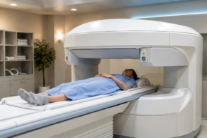
While X-rays are ideal for evaluating bones and certain chest conditions, they may not always provide enough detail for soft tissues or internal organs. If your doctor requires more detailed images, you can learn more in our MRI Scan Guide, which explains when MRI is the preferred imaging option.
How Does an X-Ray Scan Work?
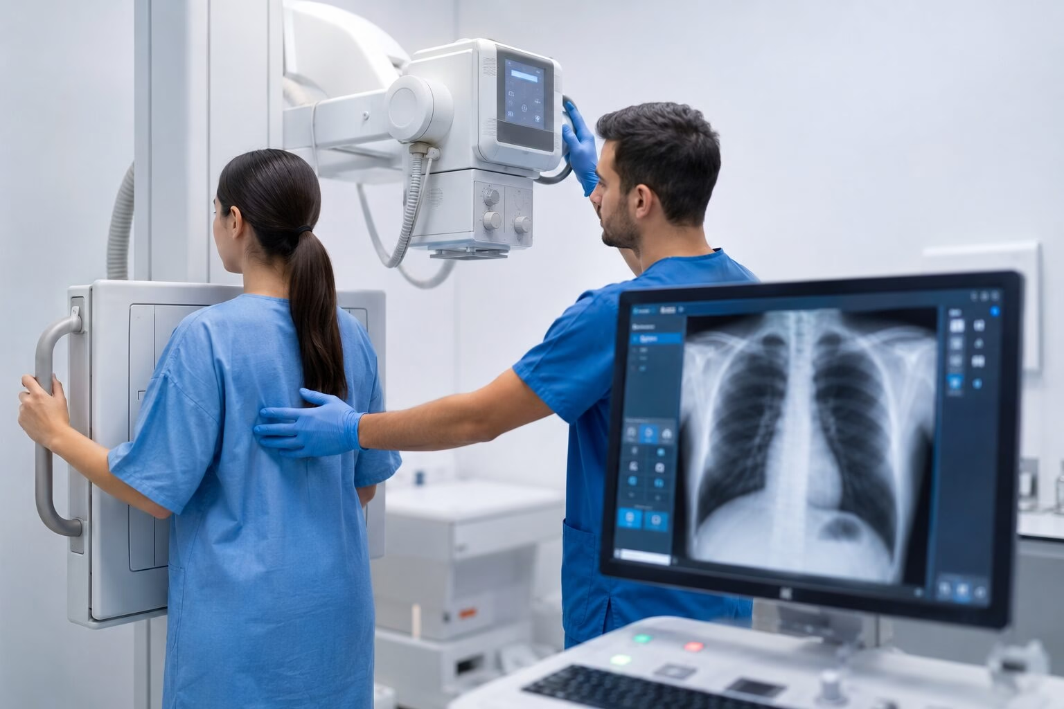
An X-ray machine produces a controlled beam of radiation that passes through the body.
As the X-rays travel through different tissues, each structure absorbs the radiation differently based on its density.
For example:
- Bones absorb most of the X-rays and appear white.
- Soft tissues absorb less radiation and appear in shades of grey.
- Air-filled spaces, such as the lungs, absorb very little radiation and appear black.
A digital detector positioned behind the body captures the X-rays that pass through and instantly converts them into detailed digital images.
The radiographer may take images from several angles to provide a more complete view of the area being examined. These images are then reviewed by a radiologist, who prepares a report for your referring doctor.
The entire process usually takes only a few minutes, making X-rays one of the quickest diagnostic imaging procedures available.
Types of X-Ray Scans
Different types of X-rays are used to examine specific areas of the body. Your doctor will recommend the most appropriate scan based on your symptoms and medical history.
Chest X-Ray
A chest X-ray is one of the most frequently performed imaging tests.
It helps evaluate:
- Lungs
- Heart
- Airways
- Ribs
- Chest cavity
Doctors commonly recommend a chest X-ray to investigate:
- Persistent cough
- Chest pain
- Shortness of breath
- Pneumonia
- Tuberculosis
- Lung infections
- Enlarged heart
- Fluid around the lungs
It is also commonly used during routine health screenings and before certain surgical procedures.
Bone X-Ray
Bone X-rays provide detailed images of the skeleton and are commonly performed following injuries or accidents.
They help diagnose:
- Bone fractures
- Stress fractures
- Bone infections
- Arthritis
- Bone tumours
- Bone deformities
Orthopaedic specialists frequently use bone X-rays to monitor healing after fractures or surgery.
Spine X-Ray
Spine X-rays evaluate the vertebrae and alignment of the spinal column.
Depending on your symptoms, your doctor may request:
- Cervical spine X-ray (neck)
- Thoracic spine X-ray (upper back)
- Lumbar spine X-ray (lower back)
These scans help identify fractures, spinal curvature, arthritis, degenerative changes, and other spinal abnormalities.
Joint X-Ray
Joint X-rays examine the bones that form major joints throughout the body.
Common joint X-rays include:
- Knee
- Shoulder
- Hip
- Elbow
- Wrist
- Hand
- Ankle
- Foot
These scans are useful for evaluating joint injuries, arthritis, dislocations, fractures, and age-related wear and tear.
Dental X-Ray
Dental X-rays allow dentists to examine teeth, jawbones, and surrounding oral structures.
Different types include:
- Bitewing X-rays
- Periapical X-rays
- Panoramic dental X-rays (OPG)
Routine dental X-rays help detect cavities, impacted teeth, gum disease, bone loss, infections, and other oral health conditions.
For more detailed three-dimensional dental imaging, your dentist may recommend a CBCT scan, which provides comprehensive 3D images for implant planning, orthodontics, and complex dental procedures.
Abdominal X-Ray
Although ultrasound and CT scans are often preferred for abdominal conditions, an abdominal X-ray remains useful in certain situations.
It may help identify:
- Bowel obstruction
- Kidney stones
- Swallowed foreign objects
- Intestinal gas patterns
- Constipation
Your doctor will determine whether an X-ray or another imaging test is more appropriate for your symptoms.
Skull X-Ray
A skull X-ray examines the bones of the skull and facial structures.
Although it has become less common due to advances in CT and MRI imaging, it may still be used in selected cases to evaluate fractures or certain bone abnormalities.
When Do You Need an X-Ray?
Your healthcare provider may recommend an X-ray whenever a quick assessment of bones, joints, or certain organs is needed.
Common reasons include:
- Bone injuries
- Suspected fractures
- Sports injuries
- Joint pain
- Persistent back or neck pain
- Chest pain
- Chronic cough
- Shortness of breath
- Swelling
- Arthritis
- Dental pain
- Suspected lung infections
- Monitoring healing after surgery or fractures
Because X-rays are fast, affordable, and widely available, they often serve as the first step in diagnosing many medical conditions.
If your symptoms involve abdominal pain, pregnancy, or blood vessel evaluation, your healthcare provider may recommend an ultrasound scan instead of an X-ray, depending on the area being examined.
X-Ray Procedure: What to Expect
An X-ray examination is simple and usually completed within a few minutes.
Step 1: Registration
After arriving at the diagnostic centre, you’ll complete the registration process and provide your doctor’s referral or relevant medical information.
Step 2: Changing Clothes
Depending on the area being examined, you may be asked to change into a hospital gown and remove clothing or accessories that could interfere with the images.
Step 3: Removing Metal Objects
You’ll need to remove jewellery, watches, belts, glasses, or other metal objects near the examination area, as these may affect image quality.
Step 4: Positioning
The radiographer will carefully position you against the digital X-ray detector.
Depending on the examination, you may be asked to:
- Stand
- Sit
- Lie down
- Raise your arms
- Hold your breath briefly
- Remain completely still
Proper positioning ensures clear and accurate images.
Step 5: Image Capture
The X-ray machine is positioned over the area being examined.
The image is captured within a fraction of a second. For some examinations, multiple images from different angles may be required.
The procedure is painless, although you may experience mild discomfort if you have an injury and need to maintain a specific position.
Step 6: Completion
Once the required images have been taken, the examination is complete.
You can immediately return to your normal daily activities unless your doctor provides additional instructions.
The images are then reviewed by a radiologist, who prepares a detailed report for your healthcare provider.
In the next section, we’ll discuss how to prepare for an X-ray, safety considerations, the differences between X-rays, CT scans, MRI scans, and ultrasound, approximate X-ray costs in Dubai, and how to understand your X-ray results.
How to Prepare for an X-Ray Scan
Most X-ray examinations require very little preparation, making them one of the most convenient diagnostic imaging tests. However, preparation may vary depending on the part of the body being examined.
General Preparation
Before your X-ray appointment:
- Wear comfortable, loose-fitting clothing.
- Remove jewellery, watches, belts, glasses, or any metal objects near the area being scanned.
- Inform the radiographer if you have any implanted medical devices or metal implants.
- Arrive a few minutes early to complete the registration process.
Contrast X-Rays
Some specialised X-ray examinations use a contrast agent to improve the visibility of certain organs or blood vessels. In these cases, your doctor may ask you to fast for several hours before the procedure or provide additional preparation instructions.
Pregnancy
If you are pregnant or think you may be pregnant, inform your healthcare provider before the examination. Although the radiation dose from a standard X-ray is low, alternative imaging methods such as ultrasound or MRI may be considered when appropriate.
Is an X-Ray Scan Safe?
Yes. X-ray scans are considered safe when performed for appropriate medical reasons.
Modern digital X-ray systems use significantly lower radiation doses than older film-based technology while producing high-quality images. Healthcare providers carefully balance the benefits of obtaining an accurate diagnosis against the minimal radiation exposure involved.
To further reduce exposure, radiographers follow strict safety protocols, including:
- Using the lowest radiation dose necessary
- Shielding other parts of the body when appropriate
- Limiting repeat scans unless medically required
- Following international radiation safety guidelines
For most patients, the benefits of an accurate diagnosis far outweigh the small risks associated with X-ray imaging.
X-Ray vs. CT Scan vs. MRI vs. Ultrasound
Different imaging techniques are designed for different medical conditions. Your doctor will recommend the most appropriate test based on your symptoms and the area being examined.
|
Feature |
X-Ray |
CT Scan |
MRI Scan |
Ultrasound |
|
Uses Radiation |
Yes (Low Dose) |
Yes |
No |
No |
|
Best For |
Bones, chest, joints |
Internal organs, trauma, complex injuries |
Brain, spine, joints, soft tissues |
Pregnancy, abdomen, blood vessels, soft tissues |
|
Image Type |
2D |
Cross-sectional |
Highly detailed soft tissue images |
Real-time imaging |
|
Scan Time |
5–10 minutes |
5–15 minutes |
20–60 minutes |
10–45 minutes |
|
Pregnancy Use |
Limited |
Usually avoided unless essential |
Often suitable when clinically appropriate |
Commonly used |
While X-rays are ideal for evaluating fractures, chest conditions, and many joint problems, your doctor may recommend other imaging tests depending on your condition. For example, an MRI scan provides greater detail for soft tissues such as the brain, spine, muscles, and ligaments, while a CT scan offers detailed cross-sectional images for trauma and complex internal conditions. Ultrasound scans are commonly preferred for pregnancy, abdominal organs, and blood flow assessments because they do not use radiation.
X-Ray Scan Cost in Dubai
The cost of an X-ray scan in Dubai depends on the body part being examined, the number of images required, and the healthcare facility.
Typical price ranges include:
|
X-Ray Type |
Approximate Cost |
|
Routine Digital X-Ray |
AED 100–250 |
|
Chest X-Ray |
AED 120–250 |
|
Bone X-Ray |
AED 120–300 |
|
Joint X-Ray |
AED 150–350 |
|
Spine X-Ray |
AED 200–400 |
|
Dental X-Ray |
AED 100–300 |
The final price may vary depending on:
- The type of examination
- The number of views required
- Whether specialist reporting is included
- Your insurance coverage
Before booking your appointment, it’s always advisable to confirm the latest pricing and insurance eligibility with your chosen diagnostic centre.
Understanding Your X-Ray Results
Once your X-ray images have been taken, they are reviewed by a qualified radiologist who prepares a detailed report for your referring doctor.
Normal Results
A normal X-ray indicates that the bones, joints, or organs examined appear healthy, with no obvious signs of injury, infection, or abnormality.
Abnormal Results
Depending on the reason for your examination, an X-ray may detect:
- Bone fractures
- Joint dislocations
- Arthritis
- Bone infections
- Lung infections such as pneumonia
- Fluid around the lungs
- Enlarged heart
- Kidney stones
- Certain bowel abnormalities
If additional detail is required, your doctor may recommend further imaging, such as a CT scan, MRI, or ultrasound.
When Will You Receive the Results?
In many cases, X-ray reports are available within 24 hours, while urgent examinations may be interpreted much sooner. Your doctor will explain the findings and discuss whether any further investigations or treatment are needed.
Where Can You Get an X-Ray Scan in Dubai?
Choosing a trusted diagnostic centre is essential for obtaining accurate images and timely results.
Look for a facility that offers:
- Modern digital X-ray equipment
- Experienced radiologists and radiographers
- Fast reporting
- Comfortable patient care
- Convenient appointment scheduling
- Comprehensive diagnostic imaging services
At Health 24/7, we provide advanced Digital X-Ray services using modern imaging technology to support accurate diagnosis across a wide range of medical conditions. Our experienced team is committed to delivering reliable imaging with patient comfort and safety as a priority.
Take the Next Step Toward Fast & Accurate Diagnosis
An X-ray scan is often the first step in diagnosing fractures, chest conditions, joint disorders, and many other health concerns. Thanks to modern digital imaging, the procedure is quick, safe, and provides valuable information that helps doctors make informed treatment decisions.
Whether you need an X-ray after an injury, to investigate persistent symptoms, or as part of a routine medical evaluation, Health 24/7 offers high-quality digital X-ray services performed by experienced professionals.
Book your Digital X-Ray at Health 24/7 today and receive fast, accurate diagnostic imaging to support your healthcare journey.
Frequently Asked Questions
How much does an X-ray scan cost in Dubai?
The cost typically ranges from AED 100 to AED 400, depending on the body part being examined and the number of images required.
Is an X-ray scan painful?
No. An X-ray is a painless procedure. You may need to remain still or hold an uncomfortable position briefly, especially if you’ve sustained an injury.
How long does an X-ray take?
Most X-ray examinations are completed within 5 to 10 minutes, although more complex studies may take slightly longer.
Is an X-ray safe?
Yes. Modern digital X-rays use a very low dose of radiation and are considered safe when performed for appropriate medical reasons.
Do I need to fast before an X-ray?
Most routine X-rays do not require fasting. However, certain contrast X-ray procedures may have specific preparation instructions.
Can I have an X-ray while pregnant?
If you are pregnant or think you may be pregnant, inform your healthcare provider before the examination. Alternative imaging methods may be recommended depending on your condition.
How soon will I receive my X-ray report?
Many diagnostic centres provide reports within 24 hours, while urgent cases may be reviewed much sooner.
What is the difference between an X-ray and an MRI?
An X-ray is primarily used to evaluate bones and certain chest conditions, while an MRI provides much more detailed images of soft tissues such as the brain, spine, muscles, ligaments, and internal organs.